Dementia: early diagnosis matters
A dementia diagnosis should not be made lightly. Among the people around the patient, it provokes about as strong a reaction as a cancer diagnosis. And yet it is important,…
News
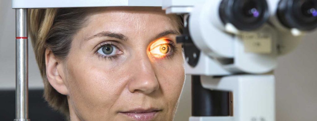
Glaucoma is a long-known chronic eye disease that can lead to blindness. The history of its name is interesting and not widely known: even the ancient Greeks noticed that some blind patients had a striking colour in their pupillary reflex. The Greek word ‘glaukos’ translates as ‘grey-bluish-greenish’. The precise anatomy of the eye and its pathophysiology were not yet understood. In the 8th century AD the term ‘grey star’ was coined for clouding of the lens, but the exact mechanism of blindness was still unclear. Only with the invention of the ophthalmoscope, which made it possible to assess the fundus and the optic nerve, could cataract and glaucoma be told apart as separate conditions.
The German term ‘Grüner Star’ (‘green star’), by contrast, was only coined in the 18th century. At the time it was believed that the ‘humours’ in the eye could change colour and would turn greenish in glaucoma. The exact origin of the term is not known, however. The disease can appear at any age, but it becomes markedly more common with age. An estimated 2.5% of all people over 40 in Switzerland have glaucoma, and, remarkably, more than half of those affected do not know it [1].
In 2004, glaucoma was the most common cause of irreversible blindness [2] and cataract the most common cause of reversible blindness. Age-related macular degeneration also causes irreversible blindness. In recent years the number of patients blinded by macular degeneration has risen sharply because of increasing life expectancy, and in some countries it has become the leading cause of blindness.
Put simply, the eyeball is a spherical structure surrounded by a firm outer layer. Each eye therefore has its own internal pressure, which is important for homeostasis. This pressure is regulated by the balance between the production and the drainage of aqueous humour. In glaucoma, the two fall out of balance, and very often it is drainage in the anterior chamber angle that is impaired. If the intraocular pressure rises above what the eye in question can tolerate, the weakest point, the optic nerve, is progressively damaged. Increasing optic nerve damage leads to progressive impairment of the visual field. It is important to stress that the optimal tolerable pressure differs from eye to eye; progressive glaucoma can also develop at a normal-tension. Current definitions of glaucoma therefore no longer treat intraocular pressure as the main factor in optic nerve damage, but as one risk factor among equals [3]. These risk factors are described in more detail in the section on diagnosis.
Glaucomas can be classified by structure or by aetiology. Structurally, there are the following forms:
By aetiology, there are two forms:
Congenital glaucomas form a subgroup and mostly belong to the primary glaucomas.
The ophthalmologist takes a history to establish the risk profile. We distinguish the following groups of risk factors [4]:
During the examination, refraction and visual acuity are measured to assess visual performance. The morphology of the eyes is assessed at the slit lamp at roughly 10 to 16 times magnification to identify any risk factors. Intraocular pressure is normally measured by Goldmann applanation tonometry (GAT), which is still the gold standard [6].
The optimal intraocular pressure lies in the range of 8–21 mm Hg. This is a statistical value, based on a mean of 15 mm Hg and the usual standard deviation.
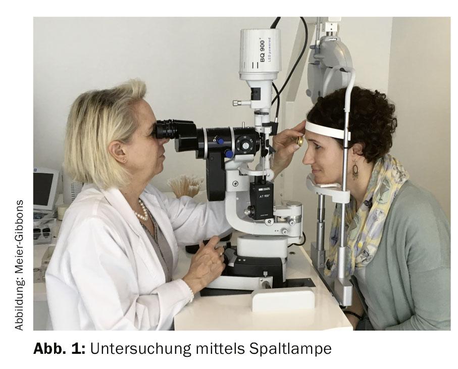
If glaucoma is suspected, further tests are ordered (Fig. 1):
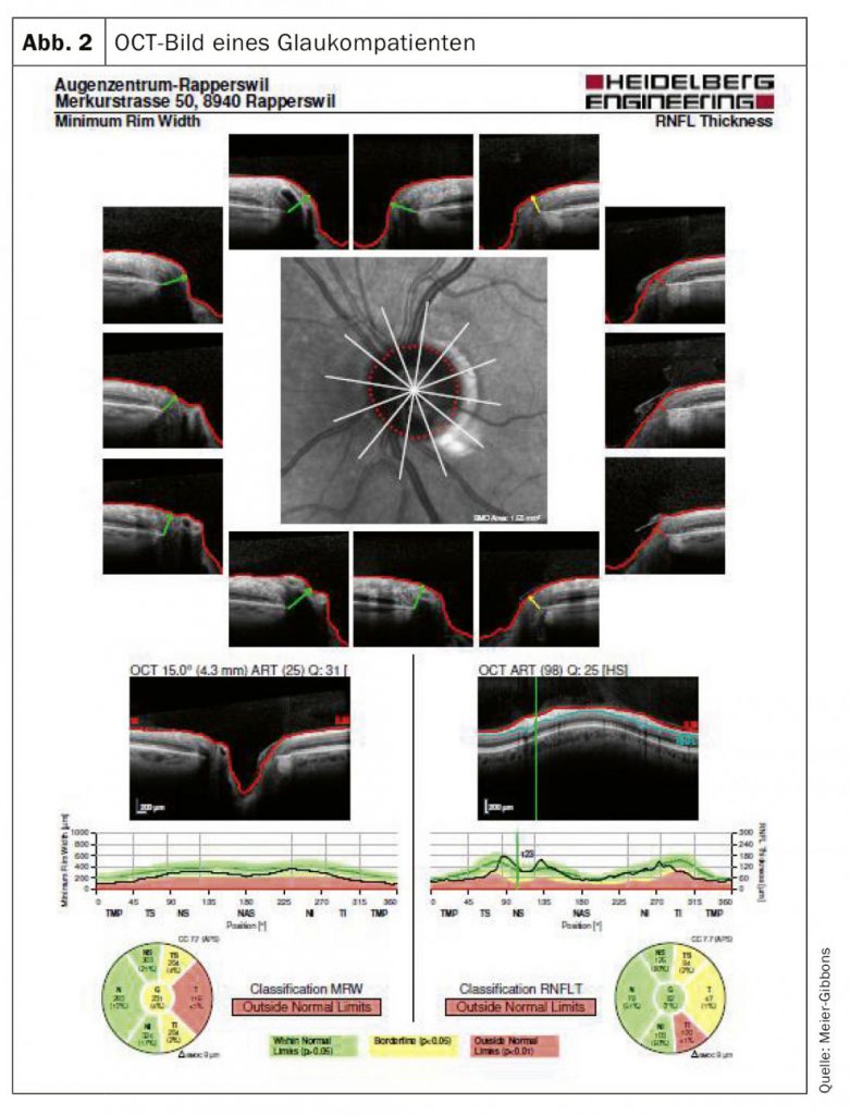
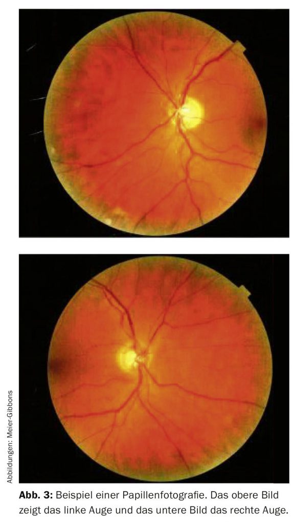
The visual field is determined by automated perimetry. In this examination, light stimuli of different sizes and intensities are presented to the patient monocularly within the normal visual field. The optic nerve should be measured in three dimensions, for which a laser-guided optical coherence tomography (OCT) scan is mostly used today (Fig. 2). This examination shows the nerve tissue layers precisely and allows them to be compared with a normative database. Colour photographs are often taken as well, since they can still be used for comparison after many years, which, unfortunately, is often not possible with OCT, because the devices are replaced so frequently (Fig. 3).
Examining the anterior chamber angle with a contact lens is important. The morphology of the angle shows what type of glaucoma is present and is important in determining treatment. The contact lens examination is non-invasive and painless for the patient.
Several newer devices for measuring intraocular pressure have been developed in recent years, because studies had shown that GAT measurement did not give the true internal pressure in certain patients (in particular with a very thin cornea, with pathological changes to the cornea or after laser correction treatment). Both these devices and corneal thickness measurement are used alongside GAT measurement.
All of the examinations mentioned above are indispensable for establishing a diagnosis. We distinguish between a normal finding, a finding suspicious for glaucoma and a clearly pathological finding.
It is important to inform the patient precisely. With a normal eye finding, a follow-up check is recommended and the patient is told that the eye can change over the course of life and that certain conditions only appear in later years. The patient must be told how often to return for checks, which depends on age, history, local findings and risk factors.
With a finding suspicious for glaucoma, informing the patient is essential: a currently borderline finding can turn out to be glaucoma within a short time. By definition (American Academy of Ophthalmology and European Glaucoma Society), a patient with suspected glaucoma has one or more of the following findings:
General note: any abnormal examination result must first be repeated to confirm it. If the repeat does show a genuine abnormality, the condition may have progressed to glaucoma.
We must remember that patients barely notice their glaucoma, at least at the start, and that every treatment has an effect and often side effects too.
The guidelines of the European Glaucoma Society state that every treatment must meet three main requirements: it must be effective and cost-effective, and cause as few side effects as possible [3].
At present, lowering intraocular pressure is the only treatment that can slow the progression of the disease. Of course, the other risk factors mentioned above must be checked and, where possible, reduced. Influencing and protecting the optic nerve itself, known as neuroprotection, would be of interest. Drug studies so far have unfortunately been disappointing.
What options do we have for lowering intraocular pressure? There are really only two:
Reducing the production of aqueous humour: aqueous humour is produced in the ciliary body, circulates in the posterior and anterior chambers of the eye and leaves the eye through the anterior chamber angle into the vascular circulation. Its production can be reduced by beta blockers (BB), alpha agonists (AA) and carbonic anhydrase inhibitors (CAI). The beta blocker timolol has been used in glaucoma treatment since 1978 and lowers intraocular pressure by 20–25%. Applied locally, BB are well tolerated, but they often cause systemic side effects such as a fall in blood pressure and pulse rate. In our view, the GP should be consulted before a patient starts BB therapy, to avoid interference with other medication.
Alpha agonists have been used for many years, but the AA available in the past often caused very marked local and systemic side effects and are no longer used. Today brimonidine is used most often. It lowers intraocular pressure by 20–25% but can also cause local (hyperaemia) and systemic (vascular and cerebral) side effects.
Carbonic anhydrase inhibitors have been commonly used systemically in glaucoma treatment since the early 1950s and have also been applied locally since 1994. They sometimes cause serious systemic side effects, particularly where there is a sulphonamide hypersensitivity. Both alpha agonists and carbonic anhydrase inhibitors are sometimes used in combination with beta blockers to achieve a better reduction in intraocular pressure.
Improving the drainage of aqueous humour: aqueous humour leaves the eye by two routes, the conventional or trabecular outflow and, much less often, the unconventional or uveoscleral outflow. In children the latter accounts for 25–55% and declines steadily with age. Reduced outflow causes fluid to back up in the eye, and intraocular pressure rises steadily. More recent studies have shown that the decisive increase in outflow resistance lies mostly in the juxtacanalicular trabecular meshwork and in the inner wall of Schlemm’s canal.
The most important drugs used to improve the drainage of aqueous humour are the topical prostaglandin agonists. They were first used in glaucoma treatment in the mid-1990s and very quickly became the leading anti-glaucoma medication. The first product was latanoprost, followed by several similar drugs. The effect (a reduction in intraocular pressure of about 25%) lasts 24 hours, which helps patients stick to treatment. Prostaglandin agonists have practically no systemic side effects, but the local side effects can be considerable: eyelash growth, increasing iris pigmentation, conjunctival hyperaemia, and also darker discolouration of the skin around the eye and atrophy of the orbital fatty tissue.
In some countries (not yet in Europe), two new groups of substances have been approved for glaucoma treatment: rho kinase inhibitors and latanoprost bunod. Both drugs improve the drainage of aqueous humour and likewise need to be applied only once a day.
It is important to us to address briefly the problem of generics, which are being used more and more often. By definition, the content of a generic should be identical to the original product. Eye drops, however, differ in essential respects from systemically administered drugs:
In our view, switching to a generic is therefore equivalent to starting a new drug and calls for more frequent check-ups at the beginning.
Pharmacies often switch to ever cheaper generics, which is problematic, because it can mean that neither the patient nor the doctor knows which drug is currently being used. It is worth discussing this with patients and pointing out the differences between original products and generics.
It is not easy to find an anti-glaucoma treatment that slows the progression of the disease, has few side effects and is used regularly. In glaucoma, as in other chronic diseases, adherence and persistence are poor. Many studies have shown that adherence to prescribed treatment is between 30% and 70%, and that after one year only 10% of patients reorder the eye drops they were prescribed [8].
There are many reasons for poor adherence, but we must remember that we are prescribing a treatment that has to be used extremely reliably and regularly and often has side effects as well. This is a particular problem for patients in the early stages of glaucoma, because they hardly notice the disease itself at this stage. In one study, Newman-Casey summarised the most common reasons for non-adherence: difficulty applying the drops, difficulty fitting the drops into the daily routine, and side effects of the drops [9]. The side effects should not be underestimated. Studies have shown that two thirds of glaucoma patients suffer side effects from their treatment [10]. Patients are bothered both by the local side effects of the drugs and by late effects on the eye, particularly on the ocular surface. If a patient has side effects from treatment, a change of treatment is worthwhile, possibly even from one prostaglandin derivative to another. In younger patients, patients on several drugs and patients who already have an ocular surface disorder (‘dry eye syndrome’), it is worth trying preservative-free eye drops. Preservatives, in particular benzalkonium chloride, can aggravate ocular surface problems. However, because preservative-free eye drops often come in single-dose packs, patients with poor visual acuity or with rheumatic changes in the hands may have difficulty applying them [11].
Besides anti-glaucoma drugs, intraocular pressure can be lowered by surgery. The first surgical procedure for glaucoma was performed in 1856 (iridectomy by Von Graefe), and the range of operations has recently widened considerably with the introduction of minimally invasive glaucoma surgery (MIGS). Various laser techniques are less invasive and can both reduce the production of aqueous humour and improve its drainage.
Surgery is considered above all in young patients with advanced glaucoma and in patients who cannot tolerate topical treatment (mostly ocular surface problems).
It is alarming that many patients still go blind from glaucoma, because its progression can clearly be influenced if the disease is detected in time and treated correctly. What do we need to work on?
At least 50% of patients still do not know that they have this disease. Precise public education is therefore essential, so that assessing patients and their risk factors can identify those with rapidly progressing glaucoma.
When treatment is started in a glaucoma patient, adherence in particular must be discussed with them: a drug applied as a drop only works if the drop is applied!
Regular checks of the optic nerve and its function are essential; intraocular pressure must be monitored and treatment adjusted accordingly. A patient diagnosed with glaucoma is usually seen by the ophthalmologist 2–3 times a year. Newer drugs with fewer side effects and possibly a different method of application (for example punctal plugs with slow release, or injection into the eye itself) can lead to better adherence and thus better control of glaucoma.
The alternatives to drug treatment are important: laser therapy can improve the drainage of aqueous humour, and surgery also offers many new approaches, with so-called ‘minimally invasive’ procedures performed more and more often, some of which involve inserting stents.
Patients will remain important in future: they must be given precise information about their disease and be involved in its treatment.
References:
HAUSARZT PRAXIS 2019; 14(11): 6–10
Dr. med. Frances Meier-Gibbons
Dr. med. Karin Øyo-Szerenyi
A dementia diagnosis should not be made lightly. Among the people around the patient, it provokes about as strong a reaction as a cancer diagnosis. And yet it is important,…
The sudden cardiac death of an apparently healthy athlete is shocking. Despite its low incidence, work is under way on ways of detecting SCD early. Among…
Hypertension, raised blood lipid levels and diabetes are among the risk factors for CHD. As primary prevention, omega-3 fatty acid supplementation and aspirin administration…